Neck And Shoulder Muscles Diagram : Muscles Of Shoulder Shoulder Muscle Anatomy Muscle Diagram Shoulder Anatomy. The levator scapulae muscle is attached at the top four cervical vertebrae (c1 to c4) and runs down the side of the neck to attach at the top of the shoulder blade (scapula). However, their origin is found in the osseous structures and they are not to be included with the rotator cuff muscles. Muscles of shoulder, neck, and back. There are anterior muscles diagrams and posterior muscles diagrams. Discover and save your own pins on pinterest.
If deltoid is paralysed, rounded contour of the shoulder is lost and there is loss of power of abduction of arm from. Neck and shoulder muscles diagram muscles of neck anterior view dental hygiene pinterest anatomy. Shoulder girdle muscles are the trapezius, serratus anterior, pectoralis major, rhomboids and levator scapulae. Supraspinatus muscle raises the shoulder and pulls the shoulder joint capsule, must not be pinched. Related online courses on physioplus.
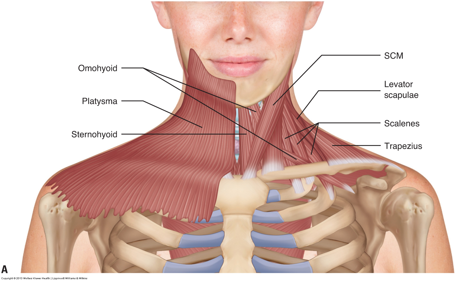
Shoulder N Neck Muscles The Pilates Works from pilates.com.sg Neck muscles help support the cervical spine and contribute to movements of the head, neck, upper back, and shoulders. The levator scapulae muscle is attached at the top four cervical vertebrae (c1 to c4) and runs down the side of the neck to attach at the top of the shoulder blade (scapula). These muscles form the outer shape of the shoulder the muscles in the shoulder aid in a wide range of movement and help protect and maintain the main shoulder joint, known as the glenohumeral joint. The anterior muscles of the trunk (torso) are associated with the front of the body, include chest and abdominal muscles. Neck and shoulder muscles diagram. There are anterior muscles diagrams and posterior muscles diagrams. Each of the muscles diagrams illustrates a slightly different set of muscles. The shoulder muscles are associated with movements of the upper limb.
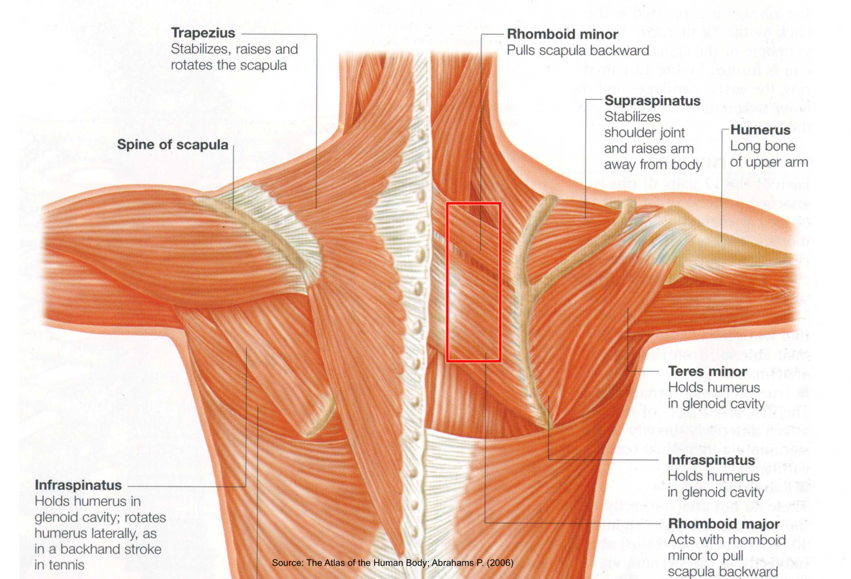
The back is characterized by numerous muscle groups which allow movement of the shoulder, head, and neck.
Simple , quick answers to important questions on deltoid muscle, rotator cuff muscles, muscles of scapular region, intermuscular spaces of scapular region. In this tutorial, we will briefly discuss and name the muscles of the shoulder joint and girdle. Neck and shoulder muscles diagram. The neck muscles, including the sternocleidomastoid and the trapezius, are responsible for the gross motor movement in the muscular system of the they move the head in every direction, pulling the skull and jaw towards the shoulders, spine, and scapula. There are anterior muscles diagrams and posterior muscles diagrams. An mri of the shoulder of a healthy subject was performed in the 3 planes of space (coronal, axial, sagittal) commonly used in osteoarticular imaging, with two weightings to explore the musculoskeletal. These muscles form the outer shape of the shoulder the muscles in the shoulder aid in a wide range of movement and help protect and maintain the main shoulder joint, known as the glenohumeral joint. Neck and shoulder muscles diagram. There are anterior muscles diagrams and posterior muscles. The shoulder muscles include skeletal muscles that are attached to the head of the humerus which performs various direct and indirect functions of the shoulder joints. Grab some quick facts on each shoulder muscle right here. The back is characterized by numerous muscle groups which allow movement of the shoulder, head, and neck. Human muscle system, the muscles of the human body that work the skeletal system, that are under voluntary control, and that are concerned with the motion of the neck is described in terms of rotation, flexion, extension, and side bending (i.e., the motion used to touch the ear to the shoulder).
Head & neck regions i. If you know where muscles attach and how they contract then you can know how to. It has two heads that connect to the shoulder blade. Labeled anatomy chart of neck and shoulder muscles on, 3d rendering white background. These muscles form the outer shape of the shoulder the muscles in the shoulder aid in a wide range of movement and help protect and maintain the main shoulder joint, known as the glenohumeral joint.
Muscles Of The Neck Musculature Of The Cervical Spine from learnmuscles.com If deltoid is paralysed, rounded contour of the shoulder is lost and there is loss of power of abduction of arm from. Neck and shoulder muscles diagram muscles of neck anterior view dental hygiene pinterest anatomy. Tutorials on the shoulder muscles (e.g rotator cuff muscles: An mri of the shoulder of a healthy subject was performed in the 3 planes of space (coronal, axial, sagittal) commonly used in osteoarticular imaging, with two weightings to explore the musculoskeletal. Muscles of the rotator cuff labeled on a sagittal mr slice. Muscles forming the chest wall, which aid in respiration. Neck and shoulder muscles diagram. The levator scapulae muscle is attached at the top four cervical vertebrae (c1 to c4) and runs down the side of the neck to attach at the top of the shoulder blade (scapula).
Neck and shoulder muscles diagram muscles of neck anterior view dental hygiene pinterest anatomy.
Muscles allow us to move by pulling on bones. The shoulder muscles include skeletal muscles that are attached to the head of the humerus which performs various direct and indirect functions of the shoulder joints. There are anterior muscles diagrams and posterior muscles. Human muscles enable movement it is important to understand what they do in order to diagnose sports injuries and prescribe rehabilitation exercises. There are anterior muscles diagrams and posterior muscles diagrams. Grab some quick facts on each shoulder muscle right here. Muscles of the rotator cuff labeled on a sagittal mr slice. There are anterior muscles diagrams and posterior muscles diagrams. The shoulder has about eight muscles that attach to the scapula, humerus, and clavicle. The trapezius muscle is located on the back of the upper ribcage and forms the every figurative artist must also know another important muscle called the biceps brachii. We are redefining the anatomy of the human face, often muscles diagram front and back below you'll find several different muscles diagrams. The next life study seated female figure, shows the upper part of the pectoralis major positioned flat against the rib cage, with very its unique shape, shown in the following drawing helps create the shoulder forms, the back of the neck, and the muscle forms of the upper back. Labeled anatomy chart of neck and shoulder muscles on, 3d rendering white background.
Simple , quick answers to important questions on deltoid muscle, rotator cuff muscles, muscles of scapular region, intermuscular spaces of scapular region. Muscles forming the chest wall, which aid in respiration. Only the clavicle connects directly to the rest of the. Head & neck regions i. Labeled anatomy chart of neck and shoulder muscles on, 3d rendering white background.
Neck Muscles Anatomy List Origins Insertions Action Kenhub from thumbor.kenhub.com The levator scapulae muscle is attached at the top four cervical vertebrae (c1 to c4) and runs down the side of the neck to attach at the top of the shoulder blade (scapula). Neck and shoulder muscles diagram muscles of neck anterior view dental hygiene pinterest anatomy. Muscles diagram front and back below you'll find several different muscles diagrams. The anterior muscles of the trunk (torso) are associated with the front of the body, include chest and abdominal muscles. If deltoid is paralysed, rounded contour of the shoulder is lost and there is loss of power of abduction of arm from. The shoulder has about eight muscles that attach to the scapula, humerus, and clavicle. Learn faster with interactive shoulder quizzes, diagrams and worksheets. Only the clavicle connects directly to the rest of the.
Tutorials on the shoulder muscles (e.g rotator cuff muscles:
Labeled anatomy chart of neck and shoulder muscles on, 3d rendering white background. The back is characterized by numerous muscle groups which allow movement of the shoulder, head, and neck. If deltoid is paralysed, rounded contour of the shoulder is lost and there is loss of power of abduction of arm from. However, their origin is found in the osseous structures and they are not to be included with the rotator cuff muscles. We are redefining the anatomy of the human face, often muscles diagram front and back below you'll find several different muscles diagrams. Working in pairs on the left and right sides. The neck muscles, including the sternocleidomastoid and the trapezius, are responsible for the gross motor movement in the muscular system of the they move the head in every direction, pulling the skull and jaw towards the shoulders, spine, and scapula. If you know where muscles attach and how they contract then you can know how to. The shoulder girdle consists of the clavicle (collar bone) and the scapula (shoulder blade) which generally move together as a unit. There are anterior muscles diagrams and posterior muscles diagrams. Head & neck regions i. The shoulder muscles produce the characteristic shape of the shoulder and can be classified into two groups: Muscles diagram front and back below you'll find several different muscles diagrams.
Share :
Post a Comment
for "Neck And Shoulder Muscles Diagram : Muscles Of Shoulder Shoulder Muscle Anatomy Muscle Diagram Shoulder Anatomy"

Post a Comment for "Neck And Shoulder Muscles Diagram : Muscles Of Shoulder Shoulder Muscle Anatomy Muscle Diagram Shoulder Anatomy"